Pain or swelling behind your last molar may make you wonder if a wisdom tooth is trapped. Impacted wisdom teeth are third molars that cannot fully erupt into the mouth because gum tissue, bone, or another tooth blocks their path. Some cause pain or infection, while others remain hidden and cause no noticeable symptoms.
An impacted tooth does not always need to be removed. The decision depends on its position, whether it can be cleaned, and whether it is harming the nearby gum, bone, or second molar. A dental examination and X-rays are needed because symptoms alone cannot show how the tooth is positioned or whether hidden damage has started.
Knowing which changes matter can help you seek care at the right time. Mild soreness behind a molar is different from increasing facial swelling, fever, or trouble swallowing. First, it helps to understand what an impacted tooth is and why some wisdom teeth cannot erupt normally.
What Are Impacted Wisdom Teeth?
Wisdom teeth are the third set of molars at the very back of the mouth. Most people develop up to four wisdom teeth, but some have fewer or none. These teeth usually try to erupt during the late teens or early adult years.
Impacted wisdom teeth cannot grow fully into the mouth because gum, bone, or another tooth blocks their path. A normally erupted tooth reaches a useful position and can usually be cleaned. An impacted tooth may remain partly visible or stay completely hidden beneath the gum and jawbone.
What Do Impacted Wisdom Teeth Look Like?
A partially impacted wisdom tooth may look like a small piece of enamel behind the second molar. It may sit under a flap of gum that becomes red or swollen. Food can also collect around this area.
Fully impacted wisdom teeth cannot be seen by looking inside the mouth. Even when part of a tooth is visible, its angle, roots, and relationship to nearby structures remain hidden. A dental examination and X-ray are needed to confirm the impaction.
Types of Impacted Wisdom Teeth
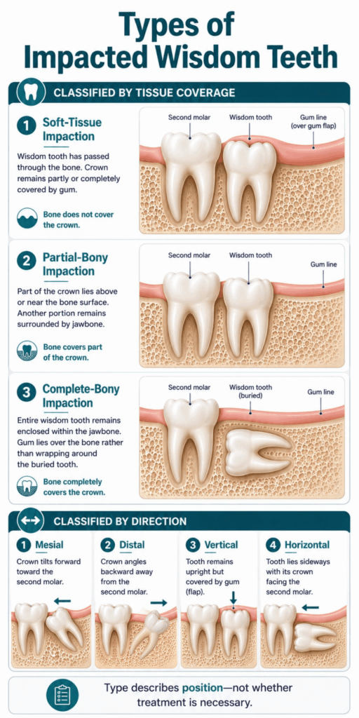
Dentists classify impaction in two main ways. The first describes how much gum or bone covers the tooth. The second describes the direction in which the tooth is positioned.
The type can affect how easily the area can be cleaned, whether the neighboring tooth is at risk, and how difficult the tooth may be to remove. However, the classification alone does not determine whether treatment is necessary.
Partial and Complete Impaction
A partially impacted tooth has broken through the gum but has not erupted fully. Part of its chewing surface may be visible, while gum still covers the rest. This can create a sheltered space where plaque, food, and bacteria collect.
A completely impacted wisdom tooth remains beneath the gum. It may be covered by soft tissue, bone, or both. Because it cannot be seen directly, a dentist usually identifies it on an X-ray.
Soft-Tissue and Bony Impaction
Dentists may describe the tissues covering the tooth:
- Soft-Tissue Impaction: The tooth has passed through the bone but remains partly or completely covered by gum tissue.
- Partial-Bony Impaction: Part of the tooth remains surrounded by jawbone, while another part lies closer to or above the bone’s surface.
- Complete-Bony Impaction: The entire tooth remains enclosed within the jawbone.
A complete-bony impaction may require a more involved surgical approach than a soft-tissue impaction. Still, symptoms, disease, root shape, age, and nearby anatomy also affect the treatment decision.
Mesial, Distal, Vertical, and Horizontal Impaction
The direction of the wisdom tooth provides another useful description:
- Mesial Impaction: The tooth tilts forward toward the second molar. This is a common position for lower wisdom teeth.
- Distal Impaction: The tooth angles backward, away from the second molar.
- Vertical Impaction: The tooth remains mostly upright but cannot erupt into a functional position.
- Horizontal Impaction: The tooth lies sideways, often with its crown facing the roots or back surface of the second molar.
A horizontally impacted tooth may contact the neighboring molar, but its position does not prove that damage has occurred. The dentist must assess the surrounding tissues and X-ray findings.
What Causes a Wisdom Tooth to Become Impacted?
Wisdom teeth become impacted when they do not have a clear path into the mouth. The jaw may lack enough room behind the second molar, another tooth may block the path, or the wisdom tooth may develop at an unfavorable angle.
Impaction is not caused by poor brushing or something the patient did wrong. Oral hygiene affects the health of an erupted area, but it does not control how the tooth develops inside the jaw. Because third molars form and move over several years, their final position may not be obvious at an early age.
Symptoms of Impacted Wisdom Teeth
Some impacted wisdom teeth cause pain or inflammation. Others cause no symptoms and are found during a routine X-ray. Symptoms are more likely when a tooth is partially erupted and difficult to clean, but even a fully impacted tooth should be assessed in context.
Pain behind the last molar does not always come from a wisdom tooth. Tooth decay, gum disease, a jaw-joint problem, and other conditions can produce similar discomfort.
Common Signs and Symptoms
Possible signs of impacted wisdom teeth include:
- Pain or Pressure: Discomfort may develop behind the last molar or along the back of the jaw.
- Tender or Swollen Gum: The tissue around a partially erupted tooth may become sore, red, or puffy.
- Food Trapping: Food may collect beneath a gum flap or between the wisdom tooth and second molar.
- Bad Breath or an Unpleasant Taste: Bacteria and trapped debris can create an odor or bad taste.
- Jaw Discomfort: Chewing may feel uncomfortable, especially near the back of the mouth.
- Limited Mouth Opening: Inflammation around a lower wisdom tooth can sometimes make it harder to open the mouth.
- Drainage: Pus or fluid near the tooth may indicate infection and requires dental care.
These findings can suggest wisdom tooth impaction, but they cannot confirm it. A dentist must examine the area and identify the actual source.
Can an Impacted Wisdom Tooth Cause Ear, Jaw, or Head Pain?
An impacted wisdom tooth may cause pain that spreads from the back of the jaw toward the ear, cheek, or side of the head. This is called referred pain. It occurs because nearby areas share connected nerve pathways.
However, ear pain and headaches can also come from an ear condition, sinus problem, jaw-joint disorder, muscle tension, or another tooth. A dental examination helps separate wisdom tooth pain from these other causes.
Can an Impacted Wisdom Tooth Cause No Pain?
Yes. A fully or partially impacted wisdom tooth may remain painless for years. Lack of pain does not always mean the tooth and surrounding tissues are disease-free.
During monitoring, a dentist checks whether the area can be cleaned and whether there is decay, gum loss, bone change, or damage to the adjacent second molar. X-rays may reveal changes that cannot be felt or seen inside the mouth.
Impacted Wisdom Tooth Pain vs. Normal Eruption Pain
Mild tenderness can occur as a wisdom tooth moves through the gum. Pain that persists, worsens, or occurs with swelling and an unpleasant taste is less likely to represent simple eruption discomfort.
| Feature | Normal Eruption Discomfort | Possible Impaction or Inflammation |
|---|---|---|
| Pain pattern | Mild, temporary tenderness | Persistent, recurring, or worsening pain |
| Location | Gum directly over the emerging tooth | Back of the mouth, jaw, or nearby areas |
| Gum appearance | Mild localized sensitivity | Redness, marked swelling, or a tender gum flap |
| Taste or breath | Usually unchanged | Bad taste, drainage, or bad breath may occur |
| Mouth opening | Usually normal | May become limited when inflammation is significant |
| Progression | Improves as the tissue settles | May return or become more intense |
Symptoms alone cannot show whether a wisdom tooth is impacted. If pain does not settle, repeatedly returns, or affects eating and mouth opening, arrange a dental examination.
Problems Impacted Wisdom Teeth May Cause
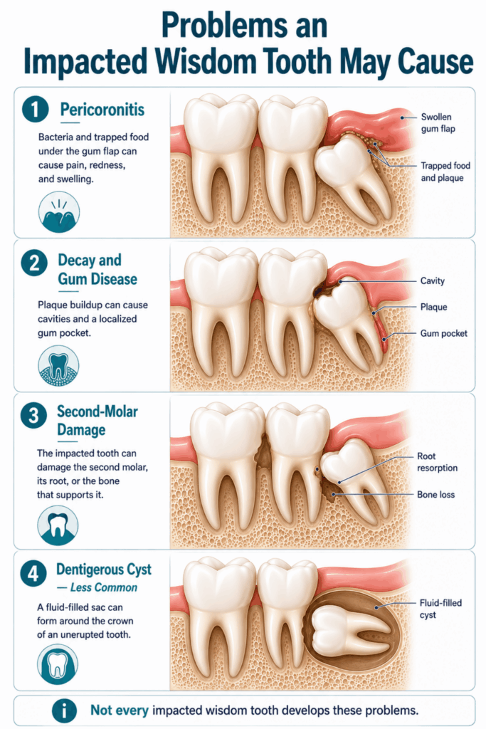
Not every impacted wisdom tooth causes disease. Risk depends on whether the tooth communicates with the mouth, can be cleaned, contacts the second molar, and remains surrounded by healthy tissues.
When problems occur, they can affect the wisdom tooth, the nearby gum and bone, or the second molar. Regular assessment matters because early damage may cause little or no pain.
Pericoronitis and Gum Infection
Pericoronitis is inflammation of the gum around a partly erupted tooth. Food and bacteria can collect beneath the gum flap, especially around a lower wisdom tooth. The area may improve and then flare again if the underlying trap remains.
Common findings include:
- Localized Pain: The gum behind the second molar feels sore or throbs.
- Redness and Swelling: The gum flap may look enlarged and feel tender.
- Bad Taste: Fluid or trapped debris may create an unpleasant taste.
- Pain When Biting: An upper tooth may press on the swollen gum below.
- Limited Opening: Increasing inflammation can make jaw movement uncomfortable.
- Spreading Symptoms: Facial swelling, fever, or difficulty swallowing requires prompt assessment.
Local cleaning and drainage are often more important than antibiotics for a localized dental infection. The Scottish Dental Clinical Effectiveness Programme’s guidance on spreading and systemic dental infection reserves antibiotics for situations such as spreading infection, systemic illness, or specific higher-risk circumstances.
Tooth Decay and Gum Disease
Partially erupted wisdom teeth can be difficult to reach with a toothbrush and floss. Plaque may remain around the tooth or beneath the gum flap, making the area prone to tooth decay and gum disease.
The second molar can also be affected. Decay may form where the two teeth meet, while persistent plaque can cause a deep gum pocket and bone loss behind the second molar. Treating these problems may be harder because access is limited.
Damage to the Nearby Second Molar
An impacted wisdom tooth may press against or sit very close to the tooth in front. The contact area can trap plaque, contribute to decay, or make cleaning nearly impossible. Gum and bone support behind the second molar may also be lost.
In some cases, pressure from the impacted tooth is associated with external root resorption, which means part of the neighboring tooth’s root is broken down. Such damage may appear on an X-ray before the patient feels pain.
Cysts and Other Less Common Complications
A fluid-filled sac called a dentigerous cyst can occasionally develop around the crown of an unerupted tooth. Other abnormal changes are much less common. These possibilities deserve attention when seen on an X-ray, but they should not be presented as an expected result of leaving every impacted tooth in place.
How Dentists Diagnose Impacted Wisdom Teeth
Diagnosis combines your symptoms, an oral examination, and dental imaging. The goal is not only to locate the wisdom tooth but also to check the gum, bone, second molar, and nearby anatomical structures.
A typical assessment includes:
- Reviewing Your Symptoms: The dentist asks when the discomfort began, whether it returns, and whether you have swelling, a bad taste, or difficulty opening your mouth.
- Examining the Area: The dentist looks for partial eruption, a gum flap, food trapping, decay, drainage, and signs of inflammation.
- Checking the Second Molar: The tooth in front is assessed for decay, gum pockets, tenderness, and possible damage.
- Taking Dental X-Rays: Imaging shows the wisdom tooth’s angle, depth, root shape, surrounding bone, and relationship to nearby teeth.
- Assessing Treatment Risk: For lower wisdom teeth, the dentist considers how close the roots appear to the nerve canal. Upper teeth are assessed in relation to nearby structures such as the maxillary sinus.
When a Panoramic X-Ray or 3D Scan May Be Used
A panoramic X-ray provides a broad view of all four wisdom teeth, both jaws, and nearby structures. It is commonly used when several wisdom teeth need assessment or surgical planning.
Cone-beam computed tomography, or CBCT, creates a three-dimensional image. It may be useful when conventional images leave a specific question unanswered, such as the relationship between a lower wisdom tooth and the inferior alveolar nerve. According to an evidence-based position paper on CBCT for mandibular third molars, it should not be used routinely before every lower wisdom tooth removal.
Do Impacted Wisdom Teeth Always Need Removal?
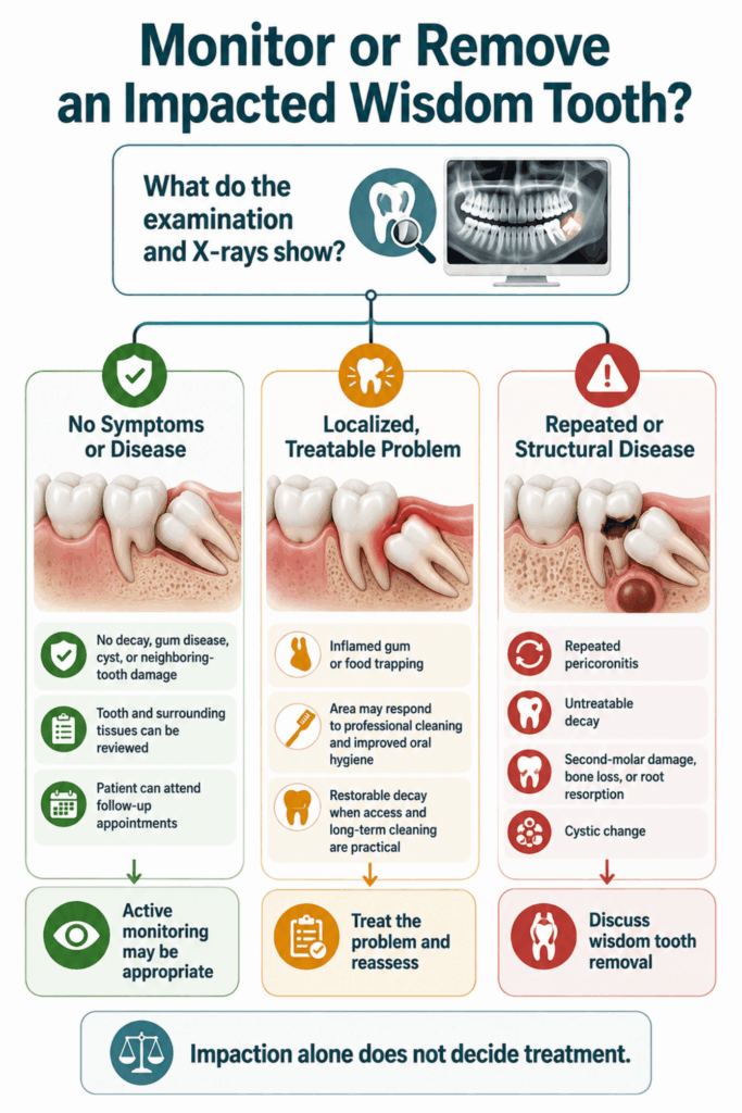
No. Impaction by itself does not mean the tooth must be extracted. Some disease-free impacted wisdom teeth can remain in place with active monitoring, while teeth causing disease or repeated symptoms may be better removed.
For symptom-free, disease-free teeth, research does not provide one answer for everyone. A Cochrane review comparing surgical removal with retention found insufficient evidence to determine whether routine removal or retention is better. The decision should consider the tooth’s health and position, cleaning access, future risk, age, general health, surgical risk, and patient preferences.
When Removal May Be Recommended
Wisdom teeth removal may be considered when the benefits are expected to outweigh the risks. Reasons can include:
- Repeated Pericoronitis: Gum inflammation keeps returning around a partially erupted tooth.
- Untreatable Decay: The wisdom tooth has decay that cannot be restored predictably because of its depth, position, or poor access.
- Damage to the Second Molar: The impacted tooth contributes to decay, gum loss, bone loss, or root resorption beside the neighboring tooth.
- Other Dental Disease: Gum disease or infection remains linked to the wisdom tooth despite appropriate care.
- Cystic Change: Imaging shows a cyst or another abnormal area associated with the tooth.
- Interference With Treatment: The tooth affects another planned procedure, such as jaw surgery or treatment of nearby disease.
The NICE recommendations on extracting impacted wisdom teeth advise limiting surgical removal to teeth with evidence of disease rather than routinely removing healthy, symptom-free impactions. Recommendations can vary by country and individual clinical circumstances.
When Monitoring May Be Appropriate
Monitoring may be reasonable when the tooth causes no symptoms, shows no evidence of disease, and is not damaging a neighboring structure. The patient must also be able to attend follow-up appointments.
Active monitoring may include:
- Scheduled Examinations: The dentist checks for new symptoms, eruption changes, or gum inflammation.
- Second-Molar Assessment: The gum, bone, and back surface of the nearby molar are reviewed carefully.
- Selected X-Rays: Imaging is repeated only when clinically justified, rather than on a fixed schedule for every patient.
- Cleaning Review: The dentist checks whether plaque and food can be controlled around any exposed part of the tooth.
- Symptom Awareness: The patient knows to report recurring pain, swelling, discharge, bad taste, or increasing difficulty opening the mouth.
Monitoring does not guarantee that the tooth will remain problem-free. It provides a way to identify changes and reconsider treatment before avoidable damage becomes extensive.
How Impacted Wisdom Teeth Are Treated
Treatment depends on what the tooth is doing now. A localized inflamed gum may need cleaning and symptom control, while repeated disease or structural damage may lead to surgical removal.
A dentist or oral and maxillofacial surgeon should identify the source before recommending care. Pain relief alone may improve symptoms without correcting the food trap, decay, or impaction behind them.
Treating Pain or Inflammation Around the Tooth
A painful area around a partially erupted tooth should be assessed, particularly if symptoms are severe or keep returning. Care may involve:
- Cleaning the Area: A dental professional may gently remove food and debris from beneath the gum flap.
- Managing the Cause: The dentist checks for biting trauma, decay, drainage needs, and other problems that could maintain the inflammation.
- Improving Oral Hygiene: Gentle cleaning around the tooth may reduce plaque buildup. Follow the dentist’s advice if the tissue is too painful to brush normally.
- Using Appropriate Pain Relief: Nonsteroidal anti-inflammatory drugs, or NSAIDs, alone or with acetaminophen, are first-line options for many people with acute dental pain, according to the American Dental Association’s guidance on oral analgesics. These medicines are not safe for everyone, so follow the label and ask a clinician or pharmacist if you have health conditions, take other medicines, are pregnant, or are unsure.
- Using Antibiotics Selectively: Antibiotics may be needed when infection is spreading, systemic symptoms are present, or particular health factors increase risk. They are not a substitute for local dental treatment.
- Planning Definitive Care: Repeated pericoronitis may lead to a discussion about removing the tooth once the acute problem is controlled.
Do not place aspirin, harsh chemicals, or unapproved home remedies on the gum. These substances can burn the tissue without treating the source.
Complete Wisdom Tooth Removal
Complete removal takes out the crown and roots. The exact procedure depends on whether the tooth is erupted, covered by bone, or close to important structures.
- Anesthesia Is Given: Local anesthesia numbs the area. Sedation or general anesthesia may also be discussed based on the procedure, medical history, anxiety, and treatment setting.
- The Tooth Is Exposed: If needed, the clinician makes a small opening in the gum.
- A Small Amount of Bone May Be Removed: This creates safe access to a bony impaction.
- The Tooth May Be Divided: Cutting the tooth into smaller sections can reduce the amount of bone removal and allow controlled extraction.
- The Area Is Cleaned: The clinician checks the tooth socket and removes loose debris.
- The Gum Is Repositioned: Stitches may be placed. Many oral stitches dissolve on their own.
Some impacted teeth are straightforward to remove, while others require maxillofacial surgery skills. Difficulty depends on factors such as depth, direction, root shape, bone density, and nearby anatomy.
Coronectomy as an Alternative to Complete Removal
A coronectomy removes the crown of a lower wisdom tooth but intentionally leaves healthy roots in the jaw. It may be considered when imaging suggests that complete root removal could place the inferior alveolar nerve at greater risk.
Coronectomy is not suitable for every tooth. Infection affecting the roots, root mobility, certain diseases, or other clinical findings may rule it out. The retained roots require follow-up because they can migrate, become exposed, or occasionally need later treatment.
What to Expect After Impacted Wisdom Tooth Removal
Some bleeding, soreness, swelling, and jaw stiffness are expected after surgical removal. The experience varies with the number and position of the teeth, the difficulty of surgery, your health, and how your body heals.
A general recovery pattern is:
- First 24 Hours: A blood clot forms in the tooth socket. Light oozing may occur, and discomfort often begins as the numbing medicine wears off. Follow the dentist’s instructions about gauze, food, medication, and cleaning.
- Days 2–3: Swelling may reach its highest point. Bruising and jaw stiffness can become more noticeable, especially after difficult lower wisdom tooth removal.
- Days 4–7: Pain and swelling should generally begin to improve. Many people return gradually to normal daily activities, although chewing may remain uncomfortable.
- Week 2: Most visible swelling and bruising have settled for many patients. The gum continues closing over the socket, and deeper healing is still underway.
- Following Weeks: Bone and soft tissue continue to remodel beneath the surface. A small depression may remain temporarily where food can collect.
Contact the treating clinic if pain becomes stronger after initially improving, bleeding will not slow, or swelling continues to increase. Follow the personal instructions given by your dentist or surgeon, since they take priority over a general timeline.
Risks of Impacted Wisdom Tooth Surgery
Wisdom tooth extraction is common, but every operation has possible complications. Your clinician should explain the risks that apply to the specific tooth before you consent.
- Dry Socket: The protective blood clot breaks down or is lost before the socket is covered by healing tissue. This can cause severe pain several days after removal.
- Infection: Increasing swelling, fever, pus, worsening pain, or a bad taste may indicate infection. Mild swelling alone during early healing does not prove that an infection is present.
- Bleeding: Mild oozing is expected at first. Persistent or heavy bleeding requires advice from the treating clinic.
- Delayed Healing: Smoking, difficult surgery, some medical conditions, and certain medicines may slow recovery.
- Nerve Disturbance: Removal of a lower wisdom tooth can irritate nearby nerves, causing altered feeling in the lower lip, chin, teeth, or tongue. Changes are often temporary, but lasting disturbance is possible.
- Damage to Nearby Structures: An adjacent tooth, filling, crown, bone, or other nearby structure can occasionally be affected.
- Sinus Communication: Upper wisdom tooth roots may lie near the maxillary sinus. A temporary opening between the socket and sinus can occasionally occur.
Risk is personal. A deeply positioned lower tooth near a nerve presents a different situation from a partly erupted upper tooth, so broad online estimates cannot replace individual assessment.
Caring for Impacted Wisdom Teeth That Are Not Removed
An impacted tooth left in place should be kept under review. When part of the tooth is exposed, careful cleaning can reduce plaque and food buildup, although it cannot change the tooth’s position.
Helpful measures include:
- Use a Small-Headed Toothbrush: Angle the bristles behind the second molar and clean the exposed surfaces gently.
- Clean Between Accessible Teeth: Floss or another dentist-recommended interdental tool may help clean the contact beside the second molar.
- Follow Individual Recall Advice: Attend dental examinations at the interval recommended for your oral health and risk level.
- Have X-Rays Only When Needed: Imaging should answer a clinical question or check a meaningful change.
- Report New Symptoms: Mention recurring soreness, swelling, drainage, bad taste, or difficulty opening the mouth.
- Avoid Cutting the Gum: Do not try to lift or trim a gum flap yourself.
- Avoid Harsh Substances: Concentrated chemicals and unapproved remedies can injure the gum.
Good oral hygiene lowers the risk of plaque-related disease, but it cannot make every partly erupted area cleanable. If inflammation keeps returning despite good care, the management plan may need to change.
When to See a Dentist
Arrange a dental appointment if pain behind the last molar persists, returns, or interferes with eating. Gum swelling, food trapping, a bad taste, drainage, bad breath from the area, or difficulty cleaning behind the second molar also deserve assessment.
Similar symptoms can come from tooth decay, a dental abscess, gum disease, jaw-joint pain, or another problem. An examination and X-rays can identify whether an impacted wisdom tooth is responsible and whether monitoring or treatment is appropriate.
When Impacted Wisdom Teeth Need Urgent Care
Most impacted wisdom tooth problems are not medical emergencies, but a spreading dental infection can become serious. Seek urgent dental or medical care if you develop:
- Rapidly Increasing Swelling: Swelling spreads across the face, below the jaw, or into the neck.
- Difficulty Swallowing: Swallowing saliva, liquids, or food becomes difficult.
- Difficulty Breathing: Breathing problems require emergency medical care immediately.
- Fever With Worsening Dental Symptoms: Fever, malaise, or feeling increasingly unwell may indicate systemic involvement.
- Severe Limitation in Mouth Opening: The jaw becomes increasingly difficult to open, especially with swelling or fever.
- Swelling Near the Eye: Upper facial swelling that approaches the eye needs prompt assessment.
Do not wait for a routine appointment if breathing or swallowing is affected. Contact emergency services or go to the nearest emergency department.
Common Questions About Impacted Wisdom Teeth
At What Age Should Impacted Wisdom Teeth Be Evaluated?
There is no single correct age for evaluation or removal. Dentists often begin assessing wisdom tooth development during the teen years, but symptoms and X-ray findings matter more than a birthday. Adults can also develop problems. Removal is considered when its expected benefits outweigh the risks, not simply because a patient reaches a certain age.
Can Impacted Wisdom Teeth Cause Your Other Teeth to Shift?
Wisdom teeth are often blamed for crowding of the lower front teeth, but tooth movement has several causes. Available evidence does not show that routinely removing disease-free impacted wisdom teeth reliably prevents orthodontic crowding or relapse. Retainers and long-term orthodontic follow-up remain important after braces.
Can You Remove Just One Impacted Wisdom Tooth?
Yes. A dentist or oral surgeon may remove only the tooth causing disease or posing a specific concern. The other wisdom teeth should still be assessed individually. Their position, health, cleaning access, and relationship to neighboring teeth help determine whether they should be monitored or treated separately.
Can a Dentist Remove an Impacted Wisdom Tooth, or Is an Oral Surgeon Needed?
Some general dentists remove straightforward impacted teeth. A referral to an oral surgeon or oral and maxillofacial surgeon may be appropriate for deep impactions, unusual roots, complex medical needs, difficult access, or close proximity to nerves and other structures. The required anesthesia or sedation may also influence the treatment setting.
Can an Impacted Wisdom Tooth Be Filled Instead of Removed?
Sometimes, but only if the decay is accessible and the tooth can be restored and cleaned afterward. A filling may not be practical when the tooth is deeply positioned, only partly erupted, repeatedly inflamed, or difficult to reach. Damage to the second molar may require separate treatment even if the wisdom tooth is removed.
Can an Impacted Wisdom Tooth Still Erupt Later?
A wisdom tooth can continue moving or erupting while the jaw and roots develop. Later eruption depends on available space, tooth angle, age, and nearby structures. An X-ray can help estimate its path, but it cannot guarantee whether the tooth will eventually reach a healthy, functional position.
Can Impacted Wisdom Teeth Affect Braces or Orthodontic Treatment?
They may influence treatment planning, especially if their position interferes with tooth movement, jaw surgery, or access to nearby teeth. However, impacted wisdom teeth do not automatically need removal before braces. The orthodontist and dentist should assess their position, health, and expected effect on the planned treatment.
The Bottom Line
Impacted wisdom teeth may cause pain, gum inflammation, decay, or damage to a neighboring tooth, but some remain symptom-free and disease-free. Impaction alone does not mean that every wisdom tooth needs removal.
A dental examination and appropriate X-rays are needed to understand the tooth’s position and health. Your dentist or oral surgeon can then compare active monitoring with treatment and help you make an informed decision based on the expected benefits and risks.
