Can you see only part of a wisdom tooth behind your last molar? Partial eruption of wisdom teeth means that a tooth has broken through the gum but has not fully emerged. It may still be moving into place, or limited space, its angle, or a gum flap may prevent it from erupting completely.
A partly erupted wisdom tooth does not always cause trouble or require removal. However, the exposed area can be difficult to clean. Food and bacteria may collect beneath the gum flap, leading to soreness, swelling, bad breath, tooth decay, or an inflamed gum condition called pericoronitis.
A dentist can examine the area and use an X-ray to check the tooth’s position, available space, and effect on the nearby molar. These findings help determine whether the tooth can be monitored, needs treatment, or should be removed. First, it helps to understand what partial eruption means and how it differs from impaction.
What Is Partial Eruption of Wisdom Teeth?
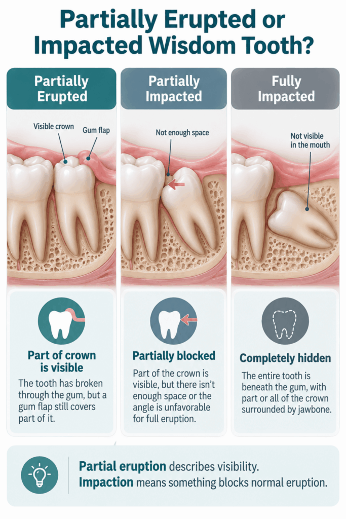
Partial eruption of wisdom teeth means that a third molar has broken through the gum tissue, but part of its crown is still covered. You may see one or more tooth cusps at the back of the mouth while the rest of the tooth remains underneath the gums.
The gum tissue covering the crown is called an operculum, or gum flap. This flap is not always inflamed or infected. However, it can create a sheltered space where plaque and food collect, especially if the tooth is difficult to reach with a toothbrush.
Is a Partially Erupted Wisdom Tooth the Same as an Impacted Tooth?
No. Partial eruption describes how much of the tooth is visible, while wisdom tooth impaction means that the tooth cannot erupt normally because something blocks its path.
A partially erupted wisdom tooth may be partially impacted, but not every partly visible tooth is permanently stuck. Some third molars continue to move into the mouth when they have enough space and a suitable angle.
| Feature | Partially Erupted Wisdom Tooth | Partially Impacted Wisdom Tooth | Fully Impacted Wisdom Tooth |
|---|---|---|---|
| Visibility | Part of the crown is visible | Part of the crown may be visible | The tooth is usually not visible |
| Position | May be upright or angled | Its position or available space prevents full eruption | It remains trapped beneath gum tissue, jawbone, or both |
| Future eruption | May continue | Less likely to erupt normally without enough space | Unlikely to erupt fully |
| Cleaning | Often difficult around the gum flap | Usually difficult because of limited access | Cannot be brushed directly |
| Possible management | Cleaning, monitoring, or treatment | Monitoring or removal, depending on disease and risk | Monitoring or surgical removal, depending on the findings |
Dentists may describe impacted wisdom teeth by the amount of soft tissue and jawbone covering them or by their angle. These types of impacted wisdom teeth include vertical, mesial, horizontal, and distal impaction. The label alone does not decide treatment; symptoms, disease, cleaning access, and effects on neighboring teeth matter more.
Why Do Wisdom Teeth Erupt Only Partway?
Wisdom teeth are the final set of molars to develop. They often begin to erupt during the late teen years or early adulthood, commonly between ages 17 and 25. By then, the other permanent teeth already occupy most of the dental arch.
Common reasons for incomplete eruption include:
- Lack of Space: There may not be enough room between the second molar and the back of the jaw for the wisdom tooth to emerge fully.
- Unfavorable Tooth Angle: A tooth angled toward the second molar, toward the back of the jaw, or sideways may become impacted instead of reaching a functional position.
- Obstruction by the Second Molar: The wisdom tooth may contact the tooth in front of it, preventing further movement.
- Gum or Bone Coverage: A tooth may emerge through the jawbone but remain partly covered by gum tissue. In other cases, part of the crown remains within both gum tissue and jawbone.
- Stage of Development: A wisdom tooth that is still developing may continue to change its angle or eruption status. A tooth that looks partially erupted today may not remain in the same position.
- Individual Anatomy: Jaw shape, tooth size, root development, and the available eruption path vary from one person to another.
A dentist cannot determine the exact reason by looking at the visible crown alone. The assessment usually includes the tooth’s angle, the space behind the second molar, and its position on an X-ray.
Can Partial Eruption of Wisdom Teeth Progress to Full Eruption?
Yes, some partially erupted wisdom teeth eventually emerge fully. The outcome depends largely on the tooth’s angle and the space available. Full eruption is more likely when the tooth is upright and moving toward a position where it can function and be cleaned.
In other cases, partial eruption of wisdom teeth persists because the tooth angle or lack of space blocks further movement. Wisdom teeth can also change position after the usual eruption age, so one examination cannot always predict their long-term course with certainty.
An X-ray helps the dentist assess whether the tooth is in the correct position for eruption. The dentist will also consider root development, bone coverage, available space, and the relationship between the wisdom tooth and second molar. Even with these details, follow-up may be needed to see whether eruption progresses.
What Does Normal Wisdom Tooth Eruption Feel Like?
A wisdom tooth that is breaking through the gum may cause mild pressure or tenderness. The area can feel different when chewing or brushing because new tooth structure is becoming exposed.
Possible features of uncomplicated eruption include:
- Mild Gum Tenderness: The gum may feel sensitive as the crown emerges.
- Temporary Pressure: You may notice a dull sensation at the back of your mouth without severe or increasing pain.
- Minor Local Swelling: A small amount of swelling may occur around the erupting tooth.
- Brief Irritation While Chewing: Food or the opposing tooth may touch tender gum tissue.
These symptoms should remain mild and improve rather than steadily worsen. Pus, marked facial swelling, fever, increasing difficulty opening your mouth, or feeling generally unwell are not expected parts of normal eruption.
Signs and Symptoms of Partial Eruption of Wisdom Teeth
Symptoms around a partially erupted tooth may come from inflamed gum tissue, infection, tooth decay, trauma from the opposing tooth, or pressure related to impaction. A symptom alone cannot identify the cause, but certain changes deserve a dental examination.
Watch for:
- Persistent or Worsening Pain: Pain that becomes stronger, throbs, interrupts sleep, or returns repeatedly may indicate more than routine eruption.
- Red or Swollen Gum Tissue: Inflammation often develops around the gum flap or behind the second molar.
- Bleeding: The area may bleed during brushing or when food irritates the tissue.
- Repeated Food Trapping: Food may collect under the flap or between the wisdom tooth and the neighboring tooth.
- Bad Taste or Discharge: A foul taste may result from trapped debris or fluid draining from an inflamed area.
- Bad Breath: Bacteria and retained food can produce an odor even when the rest of your oral hygiene is good.
- Pain When Biting or Chewing: The opposing upper tooth may press against swollen gum over a lower wisdom tooth.
- Difficulty Opening the Mouth: Jaw stiffness can develop when inflammation affects nearby muscles.
- Temperature Sensitivity: Sensitivity to cold, heat, or sweets may point to decay in the wisdom tooth or second molar.
Signs of Pericoronitis
Pericoronitis is inflammation of the gum tissue around a partially erupted tooth. It most often affects lower wisdom teeth because food and bacteria can collect beneath the operculum. The flap may become sore, red, and swollen, even if the tooth itself has no cavity.
A mild episode may cause localized tenderness, an unpleasant taste, or discomfort when chewing. More advanced infection may cause pus, tender lymph nodes, facial swelling, fever, or increasing difficulty opening the mouth. An upper wisdom tooth may make the problem worse by repeatedly biting the swollen flap over the lower tooth.
Signs of Decay or Gum Disease
Partially erupted wisdom teeth can be hard to reach with a toothbrush or floss. Plaque may remain around the wisdom tooth and along the back surface of the second molar, increasing the risk of decay and localized gum disease.
Possible signs include:
- Sensitivity: Cold, hot, sweet, or acidic foods may cause discomfort.
- Pain When Biting: A cavity or inflamed tissue may make chewing uncomfortable.
- Visible Dark Area or Hole: A mark on the tooth could represent decay, staining, or trapped debris and requires examination.
- Bleeding Gums: Inflamed gum tissue may bleed during cleaning.
- Food Trapping Between Molars: Repeated packing can irritate the gum and make plaque control difficult.
- An Unpleasant Smell or Taste: Decay and retained debris can produce odor without causing severe pain.
Decay at the back of the second molar may be difficult to see. A dentist may need an examination and appropriate dental X-rays to find it.
Problems a Partially Erupted Wisdom Tooth May Cause
Many partially erupted wisdom teeth remain stable, but their position can make them prone to oral health problems. The risk depends on whether the tooth can be cleaned, how it contacts the second molar, and whether inflammation keeps returning.
Possible problems include:
- Recurrent Pericoronitis: The gum flap may become inflamed repeatedly if bacteria and food continue to collect beneath it.
- Wisdom Tooth Decay: An erupted tooth surface can develop a cavity when brushing and fluoride exposure are limited.
- Decay in the Second Molar: An angled third molar can create a plaque trap against the back of the neighboring tooth.
- Localized Gum Disease: A deep periodontal pocket may form behind the second molar or around the partly erupted wisdom tooth.
- Damage to the Second Molar: Certain positions, especially a tooth angled forward, may be linked with decay, loss of supporting bone, or uncommon root resorption in the adjacent tooth.
- Trauma to the Gum Flap: The opposing wisdom tooth may repeatedly bite the operculum, causing soreness and swelling.
- Cystic Changes: A cyst can rarely develop around tissue associated with an unerupted or impacted tooth. Most partly erupted teeth do not develop cysts.
The goal of follow-up is to protect the entire area, not just the wisdom tooth. The Royal College of Surgeons’ third-molar care guidelines recommend assessing the eruption status, decay and periodontal health, tooth position, function, and relationship with the adjacent second molar.
How Dentists Diagnose Partial Eruption of Wisdom Teeth
A dentist combines your symptoms, a clinical examination, and imaging when needed. The aim is to determine whether the tooth is still erupting, has become impacted, or is causing disease.
- Review Your Symptoms: The dentist asks when the problem began, whether it has happened before, and whether you have swelling, discharge, fever, or difficulty opening your mouth.
- Examine the Gum Flap: The area is checked for redness, tenderness, trapped debris, bleeding, discharge, and trauma from an opposing tooth.
- Assess Cleaning Access: The dentist considers whether you can reach the tooth and whether a deep space traps plaque beneath the gum.
- Check Both Molars: The wisdom tooth and the second molar are examined for cavities, gum pockets, loss of support, and damage where the teeth meet.
- Evaluate Tooth Position and Function: An upright, functional tooth may be managed differently from one tilted against its neighbor.
- Take an X-Ray When Indicated: Imaging can show the roots, remaining bone coverage, tooth angle, available space, and relationship to nearby structures.
When a Panoramic X-Ray or 3D Scan May Be Used
A panoramic X-ray provides a broad view of all four wisdom teeth, their roots, the jaws, the second molars, and nearby structures. Dentists often use it when planning wisdom tooth removal or when a clinical finding requires a wider assessment. A smaller intraoral X-ray may be better for examining suspected decay or bone loss in a specific area.
Cone-beam computed tomography, or CBCT, produces three-dimensional images. It is not a routine test for every partially erupted or impacted wisdom tooth. According to the European Academy of DentoMaxilloFacial Radiology position paper on third-molar imaging, CBCT should be reserved for a specific clinical question that ordinary panoramic or intraoral images cannot answer. For example, an oral surgeon may consider it when the roots of a lower wisdom tooth appear closely related to the nerve canal, and the added detail could change the treatment plan.
Does a Partially Erupted Wisdom Tooth Need Removal?
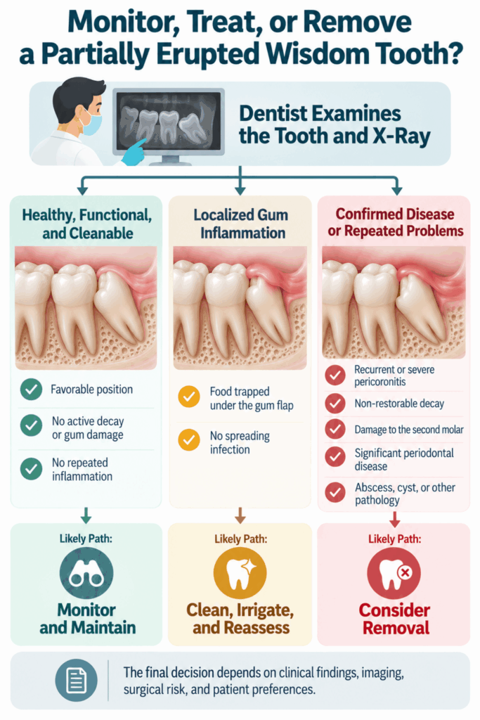
No. Partial eruption alone does not mean the tooth must be removed. The decision depends on present disease, future risk, tooth position, cleaning access, and how the tooth affects surrounding teeth.
Removal may be more likely when several of these findings are present:
- Repeated Infection: Pericoronitis returns despite appropriate local care.
- Active Decay: The wisdom tooth or neighboring second molar has a cavity that cannot be managed predictably while the third molar remains.
- Periodontal Damage: A deep pocket, inflammation, or bone loss affects the wisdom tooth or second molar.
- Unfavorable Position: The tooth is angled against the second molar or has little chance of reaching a cleanable, functional position.
- Poor Cleaning Access: Plaque and food continue to collect despite careful oral hygiene.
- Other Confirmed Disease: An abscess, cyst, or damage to a nearby tooth is present.
- Surgical Benefits Outweigh Risks: The dentist or oral surgeon decides that removal offers a better long-term outcome than continued monitoring.
A painless tooth is not automatically healthy, but it is also not automatically a reason for surgery. The choice should follow an individual assessment and a discussion of the benefits, limitations, and risks of each option.
When Monitoring May Be Reasonable
Monitoring may be suitable when the tooth is healthy, causes no active disease, has a favorable position, and can be kept clean. A partly erupted, functional third molar may sometimes be cared for like another back tooth.
Monitoring should be active rather than passive. Your dentist may check the gum, wisdom tooth, and second molar during routine visits and recommend an X-ray when symptoms or clinical findings justify it. Contact the dental office sooner if pain, swelling, food trapping, or a bad taste develops between visits.
When Removal May Be Recommended
Wisdom teeth removal may be recommended when keeping the tooth is likely to cause more harm than extraction. Common reasons include:
- Repeated or Severe Pericoronitis: A mild first episode does not always justify surgery, but repeated inflammation may support definitive treatment.
- Non-Restorable Decay: The cavity is too deep or inaccessible for a predictable filling.
- Damage to the Second Molar: The third molar contributes to decay, periodontal breakdown, or another confirmed problem affecting its neighbor.
- Periapical Infection: Infection develops around the end of the tooth’s roots.
- Cyst or Other Disease: Abnormal tissue is associated with the tooth.
- Persistent Cleaning Failure: The area cannot be maintained despite suitable home and professional care.
The NICE recommendations on wisdom tooth extraction advise against routine removal of healthy, disease-free impacted teeth. They also note that a first episode of pericoronitis, unless particularly severe, is not automatically an indication for surgery. Guidelines and professional approaches can vary, so the final decision should reflect the individual tooth and the patient’s preferences.
Treatment Options for Partial Eruption of Wisdom Teeth
Treatment depends on what is causing the problem. A dentist may first settle pain or inflammation, then decide whether the tooth can be cleaned and monitored or needs more definitive treatment.
Managing an acute episode and making a long-term decision are separate steps. Relief after cleaning or medication does not prove that an impacted tooth can erupt fully or that the problem will not return.
Cleaning and Irrigation Around the Gum Flap
A dentist may gently remove trapped food, plaque, and inflamed debris from beneath the gum flap. Irrigation flushes the sheltered space and allows the area to drain. If the upper tooth is biting the flap, the dentist will also evaluate that source of trauma.
Local treatment often helps mild pericoronitis. However, the inflammation may return if the tooth remains partly covered and the space cannot be cleaned. The recent HSE clinical guideline for pericoronitis recommends local debridement and irrigation first, with antibiotics reserved for defined signs of spreading or systemic infection.
Treating Infection and Controlling Pain
Antibiotics are not routinely required for a small, localized area of inflamed gum. Local dental treatment removes trapped debris and addresses the source. A dentist may prescribe antibiotics when infection is spreading, lymph nodes are involved, jaw opening is limited, or the patient has fever or malaise.
For temporary pain relief, adults who can safely take them may use an over-the-counter nonsteroidal anti-inflammatory drug, such as ibuprofen or naproxen, or acetaminophen. The American Dental Association’s acute dental pain guideline supports nonopioid medicines as first-line options for most acute dental pain.
Follow the product label and never exceed the stated dose. Ask a dentist, physician, or pharmacist before taking medication if you are pregnant, take blood thinners, have kidney or liver disease, have a history of stomach ulcers, have an NSAID allergy, or take medicines that may interact. Aspirin should not be placed directly on the gum because it can burn the tissue.
Removing the Gum Flap
Operculectomy is the removal or reshaping of the gum flap over a partially erupted tooth. A dentist may consider it when the tooth is upright, has enough room to erupt or function, and the flap is the main source of repeated inflammation.
The tissue can return if the tooth remains too low or lacks space to erupt. For that reason, operculectomy is not the best option for every patient. The dentist must consider tooth position, the opposing bite, cleaning access, and the chance of recurrence.
Wisdom Tooth Removal
A partly erupted tooth with enough exposed crown and a straightforward root shape may sometimes be removed with a routine extraction. The dentist loosens and lifts the tooth after numbing the area.
Surgical removal may be needed when gum tissue or jawbone still covers part of the tooth, the crown cannot be reached safely, or the roots and position make access difficult. The dentist or oral and maxillofacial surgeon may open the gum, remove a small amount of bone, or divide the tooth into sections. The recommended technique depends on the X-ray findings and nearby structures, not simply on whether the tooth is visible.
How to Clean a Partially Erupted Wisdom Tooth
When partial eruption of wisdom teeth creates hard-to-reach areas, careful cleaning helps limit plaque, food trapping, decay, and gum inflammation. Work gently because forceful cleaning can injure an already tender gum flap.
- Use a Small, Soft Toothbrush: Angle the bristles toward the back of the mouth and clean the visible crown and surrounding gumline.
- Brush the Back of the Second Molar: This surface is easy to miss and may collect plaque where it faces the wisdom tooth.
- Remove Accessible Debris Carefully: If your dentist recommends a small interdental brush, single-tufted brush, or irrigation device, follow the instructions provided. Do not force anything beneath the gum.
- Rinse After Meals: Plain water can loosen food around the tooth. A warm salt-water rinse may soothe irritated tissue, but it does not replace brushing or professional treatment.
- Use Fluoride Toothpaste: Fluoride helps protect the exposed enamel of both the wisdom tooth and neighboring molar.
- Check the Area Without Prodding It: Notice changes in swelling, color, discharge, or cleaning access, but do not squeeze the gum or attempt to drain it.
Never dig beneath the flap with toothpicks, pins, fingernails, or other sharp objects. These can damage gum tissue and push debris deeper into the area.
What You Can Do While Waiting for a Dental Visit
Home care can ease mild symptoms for a short time, but it cannot create space for an impacted tooth, remove decay, or control a spreading infection. Arrange a dental visit if symptoms persist, worsen, or keep returning.
While waiting, you can:
- Keep the Area Gently Clean: Brush the visible tooth and surrounding gum without scrubbing swollen tissue.
- Rinse With Warm Salt Water: A gentle rinse after meals may loosen food and soothe the area. Spit it out rather than swallowing it.
- Choose Foods That Do Not Pack Into the Area: Softer foods may be more comfortable during a flare-up. Avoid small seeds, sharp chips, and sticky foods that lodge beneath the flap.
- Chew on the Other Side Temporarily: This may reduce direct pressure on tender gum tissue.
- Use Pain Medicine Safely: Follow the label and confirm that the medicine is appropriate for your health conditions and current medications.
- Avoid Smoking or Vaping: Smoke and heat can irritate inflamed oral tissues and interfere with healing.
- Do Not Apply Harsh Substances: Avoid aspirin on the gum, undiluted essential oils, alcohol, bleach, or caustic home remedies.
Do not rely on leftover antibiotics. The wrong drug, dose, or treatment length may not control the source and can cause side effects or contribute to antibiotic resistance.
When to See a Dentist About a Partially Erupted Wisdom Tooth
Arrange an appointment if discomfort lasts, swelling returns, food repeatedly becomes trapped, or the area is difficult to clean. A bad taste, discharge, pain when biting, or increasing jaw stiffness also deserves assessment.
A painless, partially erupted tooth can usually be checked during a routine dental visit rather than as an emergency. Still, it should not be ignored indefinitely. The dentist can examine both molars and decide whether normal monitoring is enough.
When Symptoms Need Urgent Care
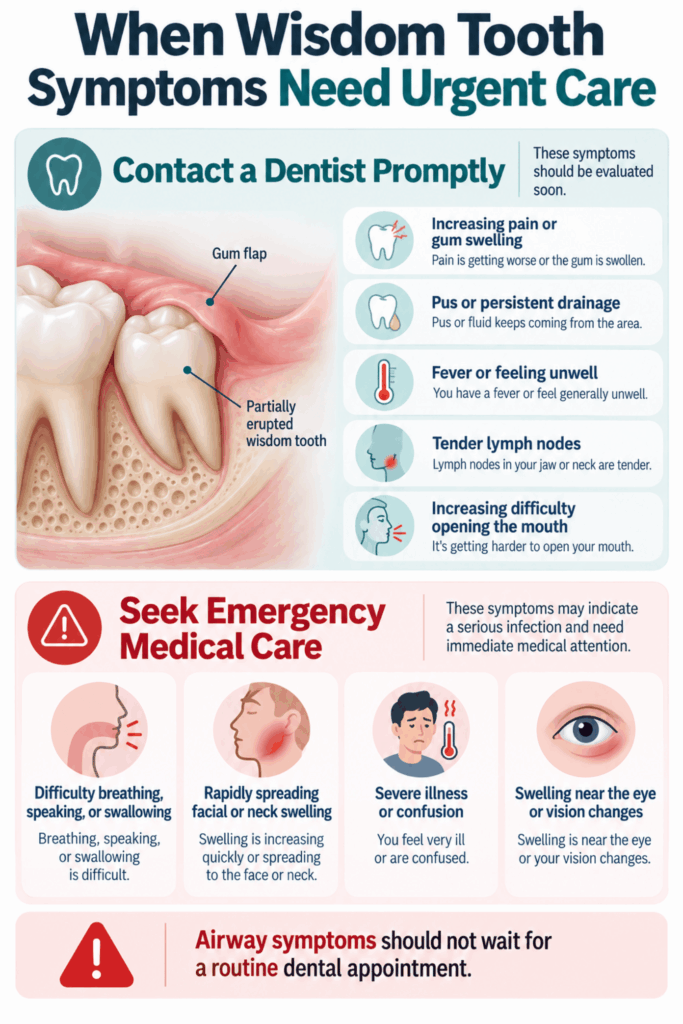
Seek urgent dental care if symptoms suggest that wisdom tooth infection is becoming more active or spreading. Contact a dentist promptly for:
- Increasing Pain or Swelling: Symptoms are becoming worse rather than settling.
- Pus or Persistent Drainage: Discharge is coming from beneath the gum flap.
- Fever or Malaise: You feel hot, weak, or generally unwell along with dental symptoms.
- Tender Neck or Jaw Lymph Nodes: Swollen glands may accompany spreading infection.
- Worsening Trismus: It is becoming difficult to open your mouth normally.
Seek emergency medical care immediately if you have:
- Difficulty Breathing or Swallowing: These can signal swelling that is affecting the airway.
- Large or Rapidly Spreading Swelling: Swelling extends into the face, jaw, floor of the mouth, or neck.
- Severe Illness or Confusion: You feel seriously unwell, or your condition is changing quickly.
- Swelling Near the Eye or Vision Changes: Infection in this area requires urgent medical assessment.
The NHS emergency guidance for dental infections advises emergency care for difficulty breathing, speaking, or swallowing and for major mouth swelling. Do not wait for a routine dental appointment when airway symptoms are present.
Can Problems Come Back After Treatment?
Yes. Pain and inflammation can return if the tooth remains partly covered and food continues to collect beneath the gum flap. Professional cleaning may settle an episode without changing the tooth’s position or improving long-term access.
Recurrence does not mean that the first treatment failed. It may show that the underlying anatomy still creates a plaque trap. The dentist will consider the number and severity of previous episodes, eruption progress, oral hygiene, tooth angle, and the health of the second molar before recommending continued monitoring, operculectomy, or tooth removal.
How to Lower the Risk of Future Problems
You cannot control the angle or available space for a wisdom tooth, but you can reduce plaque and identify disease earlier.
- Clean the Area Every Day: Use a soft brush that can reach the visible crown and the back of the second molar.
- Use Fluoride Toothpaste: This helps lower the risk of decay on exposed tooth surfaces.
- Rinse Away Loose Food: Water after meals can help when food tends to collect around the flap.
- Attend Regular Dental Checks: Your dentist can monitor the gum, wisdom tooth, and neighboring tooth.
- Report Recurring Symptoms: Repeated swelling or bad taste matters even if each episode settles.
- Follow Imaging Recommendations: Dental X-rays should be based on symptoms, examination findings, and treatment needs rather than taken automatically.
Good oral hygiene lowers risk, but it cannot make an impacted tooth change angle or create more room in the jaw. Some problems still require professional treatment despite careful cleaning.
Common Questions About Partial Eruption of Wisdom Teeth
Can a Partially Erupted Wisdom Tooth Stay Painless for Years?
Yes. A partially erupted wisdom tooth may remain painless and functional for years. However, limited cleaning access can allow decay or gum disease to develop without obvious early symptoms. Regular dental checks help monitor both the wisdom tooth and the back of the second molar.
Is a Bad Taste Around a Partially Erupted Wisdom Tooth Always an Infection?
No. A bad taste can come from trapped food, plaque, bleeding, or drainage from inflamed tissue. Infection becomes more likely when the taste occurs with increasing pain, swelling, pus, fever, or difficulty opening the mouth. A dentist must examine the area to identify the cause.
Can an Upper Wisdom Tooth Irritate the Gum Over a Lower Wisdom Tooth?
Yes. An upper wisdom tooth can bite a swollen gum flap covering a lower wisdom tooth. Repeated contact may increase pain and prevent the tissue from settling. A dentist can assess the bite and decide whether cleaning, adjustment, removal of the upper tooth, or another treatment is appropriate.
Why Does the Gum Seem to Grow Back Over My Wisdom Tooth?
The operculum may continue to cover part of the crown while eruption is incomplete. Changes in swelling can make more or less of the tooth visible, creating the impression that the gum has grown back. Gum tissue can also return after operculectomy when the tooth remains too low or lacks space.
Can a Partially Erupted Wisdom Tooth Cause Ear or Throat Pain?
Pain from an inflamed lower wisdom tooth can sometimes spread toward the ear, jaw, or throat because nearby areas share nerve pathways. However, ear and throat pain have many possible causes. Seek an examination instead of assuming that the wisdom tooth is responsible, especially if swallowing becomes difficult.
Do Both Opposing Wisdom Teeth Need Removal if Only One Causes Problems?
Not necessarily. Dentists assess each wisdom tooth separately. They may also consider whether the remaining opposing tooth will have a useful bite or could over-erupt and irritate the gum after the other tooth is removed. The decision depends on position, health, function, and surgical risk.
The Bottom Line on Partial Eruption of Wisdom Teeth
Partial eruption of wisdom teeth can have several outcomes. The tooth may continue to erupt, remain partly covered without causing disease, or create an area prone to trapped food, pericoronitis, decay, and gum problems.
A partially erupted tooth does not automatically need extraction. A dental examination can show whether cleaning and monitoring are reasonable or whether treatment would better protect your long-term oral health.
